



Ciliary sulcus IOL implantation cross section illustration | suca0255a
Price range: $19.00 through $395.00
IOL is positioned in the ciliary sulcus on top of a ruptured posterior capsule, the optic is NOT captured
- Depicts IOL placement in compromised posterior capsule
- Ideal for surgical education and patient communication
- High-resolution, ready for immediate download
- Licensed for professional and academic use
- Created by an experienced ophthalmic illustrator
✔ Royalty-free license included
✔ No Watermarks
✔ No recurring fees or attribution required
✔ Safe for professional and commercial use

Quick licensing snapshot
Enhance your ophthalmic materials with this precise ciliary sulcus IOL implant illustration.
As an eye care professional, you know how critical it is to have accurate and detailed visuals when explaining complex surgical techniques. One such technique is the implantation of an intraocular lens (IOL) into the ciliary sulcus, which is essential when the posterior capsule is compromised. This high-quality illustration of the ciliary sulcus IOL implant provides a clear and precise depiction of the technique, helping you communicate effectively in both educational and clinical settings.
In cases where the capsular bag cannot support an IOL due to rupture or damage, the ciliary sulcus becomes the alternative placement site. This illustration meticulously shows the positioning of the IOL within the ciliary sulcus, highlighting the anatomical relationships crucial for successful implantation. The clarity of this image makes it an ideal choice for ophthalmologists, educators, and researchers who need reliable visual aids.
Designed for Eye Care Professionals
Understanding surgical techniques often requires more than just textual explanations. Incorporating high-quality, realistic illustrations into presentations or educational materials enhances comprehension and engagement. This ciliary sulcus IOL implant illustration is specifically designed for ophthalmic professionals who need to demonstrate the precise placement of an IOL when the posterior capsule is compromised.
Whether you’re preparing for a surgical lecture, creating a training module, or developing patient education content, this illustration serves as a visual cornerstone. It accurately depicts the critical steps and positioning involved in IOL implantation in the ciliary sulcus, making it easier for learners and patients alike to understand the procedure.
Enhance Educational and Marketing Efforts
For medical educators, this image serves as a powerful tool to bridge the gap between theoretical knowledge and practical application. When teaching about cataract surgery complications or alternative IOL placements, this illustration makes complex concepts more accessible. Its high resolution ensures that details remain crisp, whether projected on a screen during a conference or printed in academic journals.
Marketers and content creators within the ophthalmic field will also find this illustration invaluable. Incorporating scientifically accurate visuals in presentations, websites, or educational pamphlets can significantly boost credibility. Patients and colleagues are more likely to engage with content that is both informative and visually appealing.
Why Choose This Illustration?
Many medical illustrations available online lack the necessary detail or accuracy required for professional use. I understand how crucial it is to present eye care concepts with both precision and clarity. That’s why I created this ciliary sulcus IOL implant illustration with input from experienced ophthalmic professionals, ensuring that it meets the highest standards.
The illustration is more than just visually appealing—it’s scientifically accurate. Every element, from the positioning of the IOL to the depiction of the ciliary sulcus itself, has been carefully considered to ensure that it accurately represents the surgical technique. This dedication to quality makes it a reliable resource for your professional needs.
Versatile and User-Friendly
The image file is optimized for multiple uses, making it an adaptable addition to your educational toolkit. Whether you need it for a digital presentation, a printed handout, or integration into training software, the high-resolution format ensures that the image remains sharp and clear.
Upon purchasing a license, you will have immediate access to download the file, allowing you to integrate it into your projects without delay. The licensing terms are straightforward and allow for professional, academic, and limited commercial use, giving you the flexibility to use the illustration as needed.
Real-World Applications
Surgeons who regularly perform cataract surgeries will appreciate having this illustration as part of their patient consultation process. Visual aids like this help explain why sulcus IOL implantation may be necessary when the capsular bag is compromised. By visually demonstrating the technique, patients gain a better understanding of what to expect, ultimately fostering better communication and trust.
Researchers and academic professionals can also incorporate this image into their publications or conference presentations. Accurate visuals help convey complex data more effectively, supporting arguments with visual evidence.
Invest in Quality Visual Resources
High-quality medical illustrations are an investment in professional credibility and effective communication. By choosing this ciliary sulcus IOL implant illustration, you’re ensuring that your presentations and educational materials are both accurate and visually compelling. This is not just an illustration—it’s a tool to enhance understanding and improve the quality of your ophthalmic resources.
Thank you for considering this illustration for your professional needs. I’m confident it will be a valuable addition to your collection of educational and clinical materials, supporting your efforts to deliver clear, insightful, and accurate ophthalmic content.
Similar illustrations

Phacoemulsification video clip for patient education
Phacoemulsification video clip for patient education
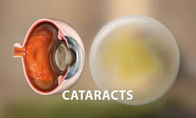
Cataract explainer video for patient education
Cataract explainer video for patient education
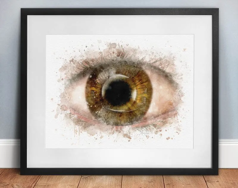
Iris fixated intraocular lens art print – 093
Iris fixated intraocular lens art print – 093

Light Adjustable Lens Implant Illustration | sui0020
Light Adjustable Lens Implant Illustration | sui0020
- 20% OFF

Cataract surgery art – step-by-step | all 4 prints for 20% off – 023c
Cataract surgery art – step-by-step | all 4 prints for 20% off – 023c

IOL art abstract wall art print eye doctor’s office décor – 092
IOL art abstract wall art print eye doctor’s office décor – 092
Join Insiders Club (free) and get 10% Off!
No spamming. Just an occasional special offer and announcements.
