



Extraocular muscles of the eye – anatomy illustration | an0002
Price range: $19.00 through $395.00
Extraocular muscles anatomy illustration: a high-quality visual for eye care professionals
- Designed for ophthalmologists, optometrists, and medical educators
- Anatomically accurate depiction of all six extraocular muscles
- Ideal for patient education, research, and professional presentations
- High-resolution download for print and digital use
- Instant licensing for seamless integration into your materials
✔ Royalty-free license included
✔ No Watermarks
✔ No recurring fees or attribution required
✔ Safe for professional and commercial use

Quick licensing snapshot
Enhance patient understanding and professional presentations with this extraocular muscles illustration
The extraocular muscles illustration is a meticulously crafted visual resource designed to clearly depict the six muscles responsible for eye movement. Whether you’re an ophthalmologist, optometrist, medical educator, or vision researcher, this high-resolution, downloadable image provides a valuable tool for explaining ocular motility, strabismus, and surgical procedures related to eye movement disorders.
Accurate anatomy for clinical and educational use
In eye care, precise anatomical visuals are crucial for effective communication. This detailed illustration ensures each extraocular muscle is correctly positioned and labeled, helping both medical professionals and patients grasp their functions. Ideal for:
- Patient consultations – help patients visualize how eye muscles work
- Medical presentations – enhance PowerPoint slides and conference talks
- Textbooks and research papers – publish with confidence using a high-quality, professional illustration
- Surgical planning – aid discussions on strabismus surgery, orbital fractures, and motility disorders
Extraocular muscles play a vital role in eye movement and binocular vision. When these muscles function properly, they allow the eyes to move together in perfect coordination, ensuring clear, stable vision. However, when one or more of these muscles are affected by injury, neurological conditions, or congenital abnormalities, eye movement can become impaired, leading to double vision, strabismus, or other visual dysfunctions. This illustration serves as an invaluable reference for understanding these disorders and guiding patient care.
Improve patient education with clear, engaging visuals
Many patients struggle to understand how extraocular muscles control eye movement. This eye anatomy illustration simplifies these complex structures, making it easier to explain binocular vision disorders, muscle imbalances, and surgical interventions. By incorporating this image into your educational materials, you can help patients feel more informed and confident in their treatment options.
Strabismus, one of the most common disorders affecting extraocular muscles, occurs when there is a misalignment in eye positioning due to weakness or overactivity in one or more muscles. This illustration provides a detailed and visually engaging way to explain different types of strabismus, such as:
- Esotropia – inward eye turn
- Exotropia – outward eye turn
- Hypertropia – upward eye deviation
- Hypotropia – downward eye deviation
Additionally, this image can assist in explaining surgical correction procedures, such as muscle recession, resection, and adjustable suture techniques, helping patients feel more confident in their treatment plan.
A must-have resource for eye care professionals
High-quality ophthalmic visuals like this extraocular muscles illustration enhance the credibility of your clinic, research, or academic materials. By using an anatomically precise and visually engaging image, you can improve information retention, patient engagement, and the overall professionalism of your presentations.
For medical educators and ophthalmic researchers, this illustration is particularly useful when discussing cranial nerve involvement in eye movement, including:
- Cranial nerve III (oculomotor nerve) – controls the superior rectus, inferior rectus, medial rectus, and inferior oblique muscles
- Cranial nerve IV (trochlear nerve) – controls the superior oblique muscle
- Cranial nerve VI (abducens nerve) – controls the lateral rectus muscle
Understanding these neural pathways is crucial for diagnosing nerve palsies, which can cause restricted eye movement and visual disturbances. This illustration is a perfect visual aid when explaining third nerve palsy, fourth nerve palsy, and sixth nerve palsy to students, residents, and patients.
Why this extraocular muscles illustration is a valuable addition to your resources
- Anatomically precise – ensures accuracy for clinical and educational use
- High-resolution quality – suitable for digital presentations, print materials, and academic publications
- Instant download – no waiting time; integrate into your work immediately
- Versatile usage – perfect for ophthalmology clinics, optometry offices, surgical guides, and medical courses
Having professionally designed, scientifically accurate visuals can make a significant difference in patient communication and medical education. This extraocular muscles illustration allows for effective knowledge transfer, helping eye care professionals communicate complex anatomical concepts in an engaging and easily understandable format.
Download and license this extraocular muscles illustration today
This high-resolution, instantly downloadable image is available with a simple licensing process, ensuring seamless integration into your medical, academic, and marketing materials. If you’re looking for a clear, accurate, and professional visual of the extraocular muscles, this illustration is designed to meet your needs. Download now and elevate your eye care resources today.
Similar illustrations

Eye drop for AMD – macular degeneration | suvr0019a
Eye drop for AMD – macular degeneration | suvr0019a

Eye anatomy watercolor art print for eye care office decor
Eye anatomy watercolor art print for eye care office decor
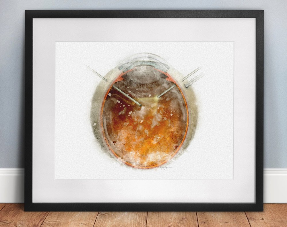
Pars plana vitrectomy art print – 184
Pars plana vitrectomy art print – 184
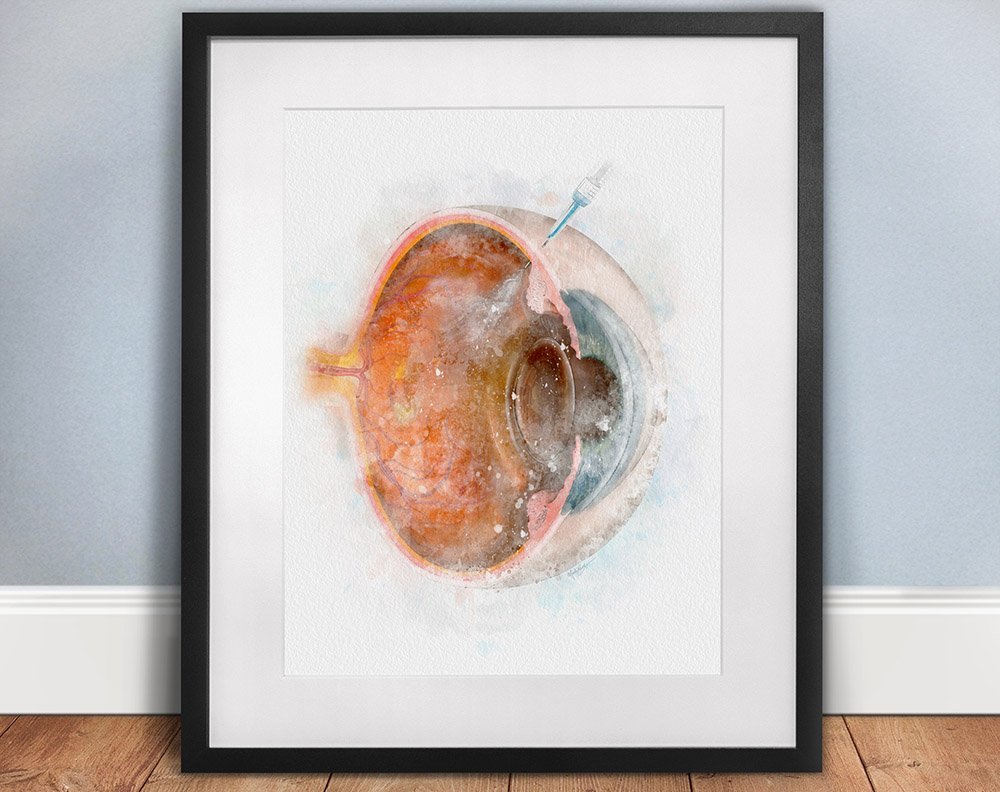
Intravitreal injection ophthalmology art print for eye care clinics – 088
Intravitreal injection ophthalmology art print for eye care clinics – 088
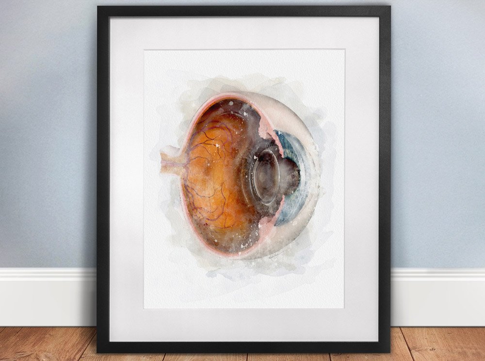
Eye anatomy art print for optometry and ophthalmology professionals
Eye anatomy art print for optometry and ophthalmology professionals

Eye injection sites and techniques, ophthalmology illustration | suvr3018
Eye injection sites and techniques, ophthalmology illustration | suvr3018
Join Insiders Club (free) and get 10% Off!
No spamming. Just an occasional special offer and announcements.
