


Browse Eye Images by Category

Eye chart for checking visual acuity | e0050

Eye chart with macular degeneration vision distortion effects | e0053

Laser peripheral iridotomy (LPI) for glaucoma | sug0060

Fluorescein angiography and OCT combined graphic | cotfa021bw

Optic nerve cupping progression in glaucoma | co3611

Glaucoma optic nerve 0.9 c/d cupping | co3610

Glaucoma optic nerve 0.8 c/d cupping | co3608

Glaucoma optic nerve 0.6 c/d cupping | co3606

Optic nerve illustration file for professional ophthalmic use | an3035

Proliferative vitreoretinopathy (PVR), retinal detachment | co3590

Exudative macular degeneration illustration | co3530a

Wet macular degeneration illustration | co3530

Coates disease retinal disorder eye illustration | co3525

Cystoid macular edema (CME) patient illustration | co3515

Cystoid macular edema (CME) fundus illustration | co3515a

Slit lamp exam on a macular hole, illustrated | co3510a

Macular hole slit lamp exam illustrated | co3510

Diabetic retinopathy findings schematic illustration | co3501

Central serous chorioretinopathy fundus image DOWNLOAD

Central serous chorioretinopathy image DOWNLOAD

Leber congenital amaurosis (LCA) | co3490a

Leber congenital amaurosis (LCA) | co3490

Amelanotic eyelid melanoma image DOWNLOAD
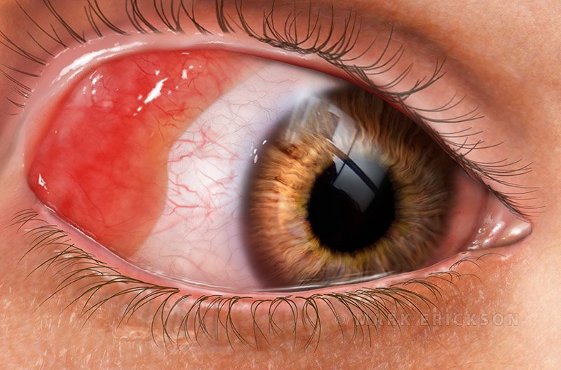
Conjunctival Lymphoma Medical Eye Illustration | co3479

Squamous cell conjunctival carcinoma illustration | co3475

Iris melanoma uveal tumor slit lamp illustration | co3466a

Iris melanoma uveal tumor illustration | co3466
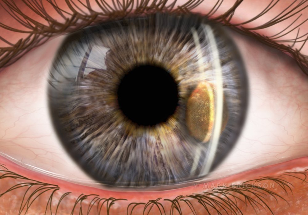
Iris melanoma slit lamp exam illustration | co3465a
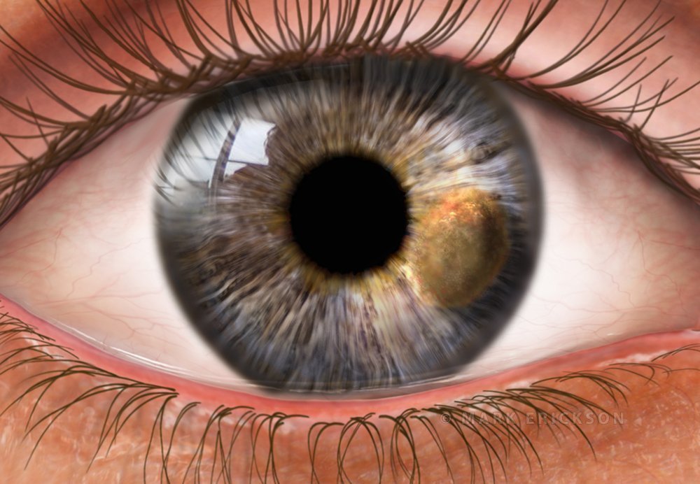
Iris melanoma illustration | co3465

Choroidal melanoma cross section illustration | co3460

Choroidal Melanoma Medical Illustration for Patient Education | co3458
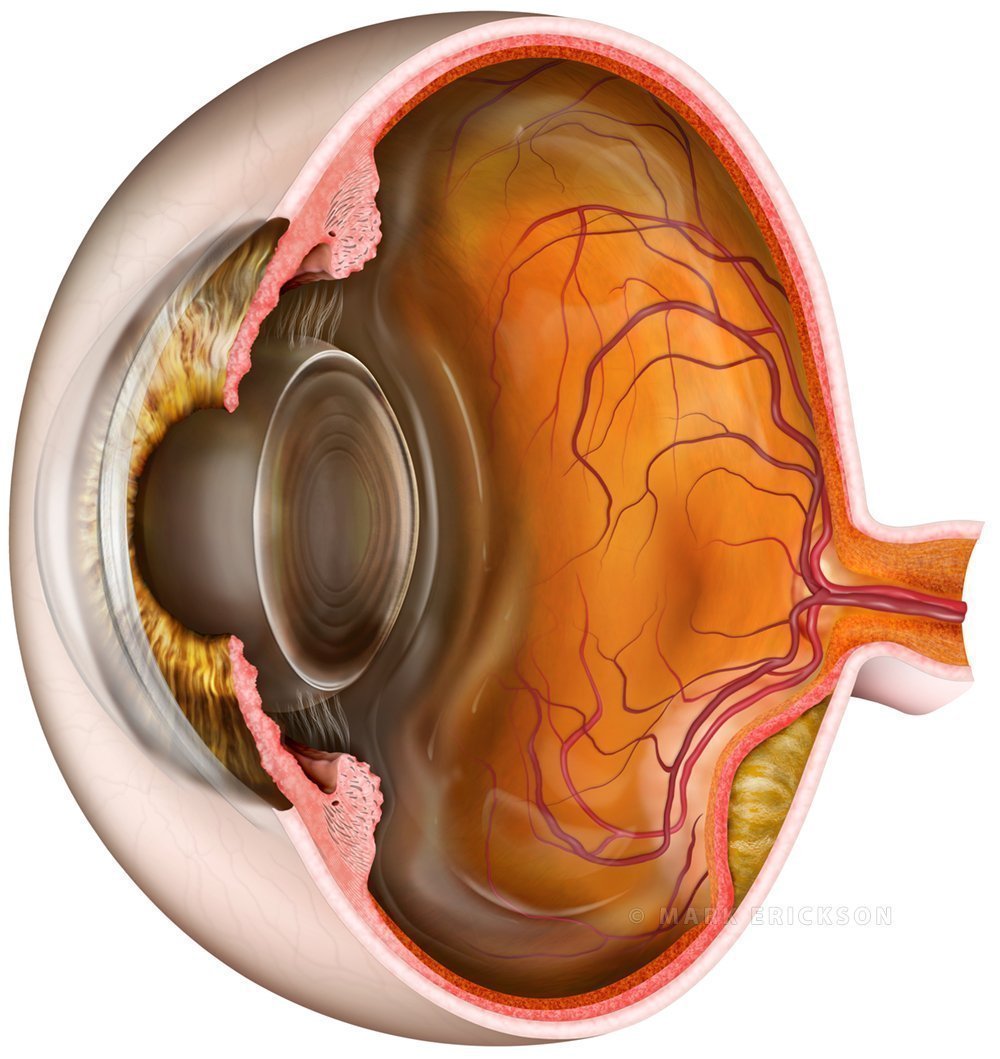
Amelanotic choroidal melanoma eye tumor cross section DOWNLOAD
